Ryle's Tube, NG Tube & Feeding Tubes: Complete Guide
Need expert consultation? Book an appointment with Dr. Vidhyadharan at THANC Hospital.
Book AppointmentFeeding tubes are one of the most common interventions in hospital care, yet they are also one of the most anxiety-inducing for patients and families. Whether it's a Ryle's tube placed in the emergency room after a stroke, a nasogastric tube inserted during cancer treatment, or a PEG tube recommended for long-term nutrition, families are often left with more questions than answers. How long will this be needed? Can my loved one ever eat again? How do we manage this at home?
At THANC Hospital in Kilpauk, Chennai, I see these questions daily. Many families arrive with the feeding tube already in place, often placed at a referring hospital weeks earlier, but without a clear plan for what comes next. The tube is keeping the patient nourished, but nobody has assessed whether the swallowing problem is improving, stable, or getting worse. This gap between tube placement and swallowing rehabilitation is where patients lose valuable recovery time.
This guide explains everything patients and families need to know about feeding tubes: what types exist, when they are needed, how to care for them, and most importantly, the path toward removing them.
Types of Feeding Tubes
Ryle's Tube (Nasogastric Tube / NG Tube)
The Ryle's tube: named after British physician Dr. John Alfred Ryle, is the most common type of feeding tube used in Indian hospitals. In medical terminology, it is called a nasogastric (NG) tube.
What it is: A thin, flexible polyurethane or silicone tube, typically 80-120 cm long, inserted through the nostril, down the pharynx, through the esophagus, and into the stomach.
When it is used:
- Acute stroke with unsafe swallowing
- Post-operative patients who cannot eat temporarily
- Head injury patients with reduced consciousness
- Cancer patients during treatment
- Any condition causing temporary inability to swallow safely
Advantages:
- Non-surgical, placed at the bedside in minutes
- No special equipment needed for placement
- Easily removable when no longer needed
- Can be replaced if dislodged
Limitations:
- Uncomfortable for long-term use
- Visible on the face (cosmetic concern)
- Can cause nasal irritation, sinusitis
- Risk of accidental dislodgement
- Must be replaced every 4-6 weeks
- Higher aspiration risk compared to PEG tubes (if tube migrates)
PEG Tube (Percutaneous Endoscopic Gastrostomy)
What it is: A tube placed directly through the abdominal wall into the stomach, using endoscopic guidance. The external portion is a small, flat disc against the abdomen with a tube connector for feeding.
When it is used:
- When tube feeding is needed for more than 4-6 weeks
- Patients with chronic swallowing disorders (stroke, TBI, neurological disease)
- Head and neck cancer patients undergoing prolonged treatment
- When patient comfort and quality of life are prioritized
Advantages:
- More comfortable than an NG tube for long-term use
- Not visible on the face
- Lower risk of sinusitis and esophageal irritation
- Lower risk of accidental dislodgement
- Easier for home care and self-management
- Can last 1-2 years before needing replacement
Limitations:
- Requires a minor endoscopic procedure for placement
- Small risk of surgical complications (infection, bleeding, leakage)
- Requires wound care around the insertion site
- Not easily reversible (though the tract heals after removal)
Other Feeding Tube Types
| Type | Route | Duration | Typical Use |
|---|---|---|---|
| Nasojejunal (NJ) tube | Nose → jejunum (small intestine) | Short-term | Patients with gastroparesis or severe reflux |
| Gastrostomy button | Abdominal wall → stomach | Long-term | Low-profile alternative to PEG for active patients |
| Jejunostomy (J) tube | Abdominal wall → jejunum | Long-term | Post-gastric surgery or severe gastroparesis |
When Is a Feeding Tube Needed?
The decision to place a feeding tube is based on two key assessments:
1. Swallowing Safety Assessment
Using FEES (Flexible Endoscopic Evaluation of Swallowing), Dr. Vidhyadharan determines whether the patient can swallow safely. If FEES shows:
- Aspiration (food or liquid entering the airway), especially silent aspiration
- Severe pharyngeal residue (large amounts of food remaining in the throat after swallowing)
- No safe food texture: even pureed food or thickened liquids are aspirated
Then oral feeding is unsafe, and a feeding tube becomes medically necessary to prevent aspiration pneumonia.
2. Nutritional Adequacy Assessment
Even if swallowing is partially safe, a feeding tube may be needed if the patient:
- Cannot eat enough calories or drink enough fluids by mouth
- Is losing weight despite oral feeding
- Takes excessively long to eat (mealtime fatigue)
- Cannot meet medication delivery requirements orally
In these cases, the feeding tube supplements oral intake rather than replacing it entirely.
Have questions about your condition? For personalized treatment options and expert care, consult Dr. Vidhyadharan Sivakumar at THANC Hospital. Call +91 73059 53378 or book an appointment.
Ryle's Tube Care: A Practical Guide for Families
Managing a Ryle's tube at home requires knowledge and confidence. Here is what families need to know.
Daily Care Checklist
- Tube position check: Before each feed, gently aspirate with a syringe to confirm gastric contents (acidic, green-yellow fluid). If in doubt, do not feed and contact the medical team.
- Nasal care: Clean the nostril around the tube daily with saline-soaked gauze. Alternate nostrils if the tube needs replacement.
- Tape security: Check that the tube is securely taped to the nose and cheek. Replace tape if loose.
- Flushing: Flush the tube with 20-30 ml of warm water before and after each feed, and before and after each medication.
- Tube patency: If the tube feels blocked, try gentle flushing with warm water. Never force a blocked tube.
Feeding Through a Ryle's Tube
- Position: Sit the patient upright (30-45 degree head elevation) before, during, and for 30-60 minutes after feeding
- Rate: Administer feeds slowly (over 20-30 minutes for bolus feeds). Gravity feeding is preferred over syringe pushing.
- Volume: Follow the dietitian's prescribed volume and frequency. Do not increase without medical advice.
- Temperature: Feeds should be at room temperature. Cold feeds can cause cramping.
- Hygiene: Wash hands before handling the tube. Use clean syringes and containers.
When to Seek Medical Help
Contact THANC Hospital or your treating team if:
- The tube comes out (do NOT attempt reinsertion at home)
- The patient develops vomiting, abdominal distension, or severe discomfort
- The patient develops fever, cough, or breathing difficulty (possible aspiration)
- The tube appears blocked despite gentle flushing
- Blood is visible in the tube or around the nostril
- The patient's nose shows signs of skin breakdown or ulceration
The Path to Tube Removal: FEES-Guided Rehabilitation
A feeding tube should never be a permanent solution unless specifically indicated. The goal is always to work toward safe oral feeding. At THANC Hospital, Dr. Vidhyadharan follows a structured pathway.
Step 1: FEES Assessment of Current Swallowing
- Evaluate what the patient can and cannot swallow safely
- Identify specific swallowing deficits (aspiration, residue, timing)
- Determine if any compensatory strategies improve safety
Step 2: Targeted Swallowing Therapy
- Exercises to strengthen weakened muscles, tongue exercises, effortful swallow, Mendelsohn maneuver
- Sensory stimulation to improve swallowing reflexes
- Compensatory strategies: chin tuck, head rotation, multiple swallows
Step 3: Graduated Oral Feeding Trials
- Begin with the safest texture identified on FEES (usually pureed foods or thickened liquids)
- Gradually increase oral intake volume while monitoring for aspiration signs
- Supplement with tube feeding to maintain nutrition during the transition
Step 4: Tube Feed Reduction
- As oral intake increases, tube feeds are reduced proportionally
- Regular weight monitoring to ensure nutritional adequacy
- Serial FEES assessments to confirm ongoing safety
Step 5: Tube Removal
- When oral intake consistently meets caloric and hydration needs for several consecutive days
- Final FEES confirms safe swallowing across required textures
- Patient maintains weight on oral diet alone
Read our detailed guide on feeding tube removal after cancer treatment.
Special Considerations
Feeding Tubes After Stroke
Post-stroke patients are the most common group requiring feeding tubes due to dysphagia. Many stroke patients can regain swallowing with structured rehabilitation. The key is not to wait passively, active swallowing therapy should begin while the patient is still tube-fed. Read more about post-stroke dysphagia rehabilitation.
Feeding Tubes After Head Injury
TBI patients often require both a tracheostomy tube and a Ryle's tube. Managing the path to removing both requires systematic FEES-guided rehabilitation. Read more about TBI swallowing rehabilitation.
Feeding Tubes in Cancer Patients
Head and neck cancer treatment (surgery and radiation) frequently causes swallowing dysfunction requiring feeding tube support. Many cancer patients can eventually return to oral eating with rehabilitation. Read more about cancer swallowing rehabilitation.
Feeding Tubes in Elderly Patients
The decision to place a feeding tube in elderly patients, especially those with dementia, requires careful ethical and medical consideration. Read more about geriatric swallowing disorders.
Why Choose Dr. Vidhyadharan for Feeding Tube Management?
- MCh (Head & Neck Surgery), Amrita Institute
- MS (Otorhinolaryngology), Gold Medal, First Mark, Annamalai University
- 15+ years specialist practice | 3000+ surgeries
- FEES-guided tube-to-oral feeding rehabilitation
- 80% decannulation success rate for eligible patients
- Structured tube weaning protocols
- Comprehensive caregiver training program
View Dr. Vidhyadharan's full academic profile and publications.
Conclusion
A feeding tube is a bridge, not a destination. Whether it's a Ryle's tube placed after a stroke, a PEG tube during cancer treatment, or a nasogastric tube after a head injury, the goal is always to work toward the safest and most functional feeding method for each patient. Families should not accept "wait and see" as a management plan. Structured swallowing assessment and rehabilitation, guided by FEES, gives the best chance of returning to oral eating and removing the tube.
If your loved one has a feeding tube and you don't have a clear plan for swallowing rehabilitation, it's time to seek specialized evaluation. Early intervention leads to better outcomes.
For personalized treatment options and expert care, consult Dr. Vidhyadharan Sivakumar at THANC Hospital, Kilpauk, Chennai. Call +91 73059 53378 or book an appointment.
Related Resources
Authored by
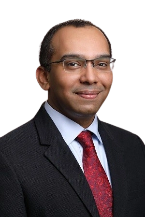
Dr. Vidhyadharan Sivakumar
MCh (Head & Neck Surgery), FEB-ORL HNS, MS (ENT) Gold Medal
Clinical Director & Senior Consultant at THANC Hospital, Chennai. Co-Editor of "Comprehensive Management of Head and Neck Cancer" (2021) with 40+ publications. Team Leader for India's first TORS-assisted Total Laryngectomy (2022). 15+ years experience with over 3000 complex surgeries.